A web site full of stuff that should be useful
| AS Psychology |
| Core Studies |
| Links |
| Course Content |
| Exam Questions |
| Psychological Investigation |
| Themes and Perspectives |
| Glossary |
| About this site |
| Forum |
| For Teachers |
|
|
Non
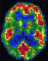
NGRI PET scan

NGRI PET scan
The two PET scans show less activity (red regions) in the prefrontal cortex of a NGRI brain
![]()
The Raine Page
|
Below is a very brief summary of the Raine study. You will need to use the more detailed summary here to revise for the exam. You can also find all of the past exam questions on Raine's study here. One of Adrian Raine's websites is here There are many web sites showing good images of PET scans. Here is one.
|
Adrian Raine
|
|
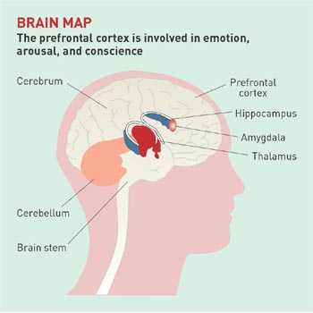
The aim of the experiment was to discover if murderers who have pleaded not guilty by reason of insanity (NGRI) show evidence of brain abnormalities. The study used PET scans to examine the brains of 41 people (39 males and 2 females) who were charged with murder and were pleading Not Guilty for Reasons of Insanity (NGRI), and compared them with 41 controls. All the NGRIs were referred to the imaging centre for legal reasons, such as to obtain evidence for the defence. The reasons for the referrals included schizophrenia, head injury, and personality disorders. The participants were matched by age and sex to a control group of participants. The participants with schizophrenia were matched with other people with the same diagnosis but no history of murder. All offenders were in
custody and were kept medication free for the two weeks before brain
scanning. The control group were also medication free. All of the participants
were injected with a glucose tracer, required to work at a continuous
performance task that was based around target recognition for 32 minutes,
and then given a PET scan. The NGRIs were compared with the controls on the
level of activity (glucose metabolism) in right and left hemispheres of the
brain in 14 selected areas. The researchers looked at activity in six
cortical areas (part of the cerebral cortex which is the outermost layer of
nerve tissues of the cerebral hemispheres) and eight subcortical areas
(brain structures below the cortex); The cerebral cortex is
commonly described in terms of four areas or lobes; the prefrontal,
parietal, temporal, and occipital. In this study, compared to the
controls, the NGRIs were found to have less activity in their prefrontal and
parietal areas, more activity in their occipital areas, and no difference in
their temporal areas. The results from the subcortical areas found less activity in the corpus callosum They also found an imbalance of activity between the two hemispheres in three other subcortical structures. In the amygdala and the hippocampus, compared to the controls, the NGRIs had less activity in the left side and more activity in the right side. Also, in the thalamus the NGRIs had more activity in the right side, though no difference in the left side. Raine et al. argue that
the difference in activity in the amygdala (which is part of the limbic
system) can be seen to support theories of violence that suggest it is due
to unusual emotional responses such as lack of fear. The authors also
comment on the differences in corpus callosum activity between the NGRIs and
the controls, and suggest this can be matched up to evidence of people with
a severed corpus callosum which show they can have inappropriate emotional
expression and an inability to grasp long-term implications of a situation. It is important to note
that Raine et al. are cautious about the implications of their findings.
There is evidence to suggest that murderers pleading NGRI have significantly
different levels of activity in the brain and that these differences may
predispose such individuals towards violence. However the researchers note
that these findings should not be taken to indicate that violence is
determined by biology alone. |
|
|
|
|