|
Background
Human brains are divided into hemispheres –
a left hemisphere and a right hemisphere.
Although these halves look similar they have very different
functions. This
division of tasks between the hemispheres is called lateralisation
of function. For
example, the major functions of language are carried out in the left
hemisphere (for right handed people).
The table below gives a summary of some of the
functions you need to know for this study.
|
Function or Stimulus
|
Part of the Brain
|
|
The right visual field
|
Left hemisphere
|
|
The left visual field
|
Right hemisphere
|
|
Language
|
Left hemisphere
|
|
Information from the left hand
|
Right hemisphere
|
|
Information from the right hand
|
Left hemisphere
|
|
Information from the left ear
|
90% right hemisphere
|
|
Information from the right ear
|
90% left hemisphere
|
|
Information from the left nostril
|
Left hemisphere
|
|
Information from the right nostril
|
Right hemisphere
|
The hemispheres are joined at their base by
commissural fibres, which allow the hemispheres to communicate
internally. The
commissural fibres are bundled into structures called commissures
(these include the corpus callosum).
These fibres are sometimes cut by surgeons to
reduce the effects of epilepsy.
Epileptic seizures are caused by millions of brain cells
firing excessively. When
someone has a grand mal (major epileptic seizure) they can have
uncontrollable movements and eventually a loss of consciousness.
Many of these seizures can involve both hemispheres and it
was noticed by surgeons that if the hemispheres were separated the
seizures could be contained in one half of the brain, therefore
causing less damage to the person.
Such surgery however was only used as a last resort if
medication could not control the epilepsy.
Roger Sperry was able to take the opportunity
of studying patients with hemisphere deconnection in order to
determine whether there are differences between the two hemispheres
of human brains.
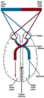
The table above points out that information
from the left visual field is processed in the right hemisphere and
vice versa. The
diagram below shows how this works.
If a person is looking straightforward
everything to the left of their nose is the left visual field and
everything to the right is their right visual field.
Everything in the persons left visual field is received to
the right of their retina and then via the optic chiasma the
information goes to the right hemisphere.
Visual Pathways of the Brain
Aim
The aim of this study was to investigate the
effects of hemisphere deconnection and to show that each hemisphere
has different functions.
Method/Procedure
The participants were 11 ‘split-brain’
patients, that is, they were patients who had undergone
disconnection of the cerebral hemispheres.
The participants had all undergone hemisphere deconnection
because they had a history of advanced epilepsy which could not be
controlled by medication.
The method used was a natural (also called
quasi) experiment. The quasi-experiments involved comparing the
performance of the 11 participants on various tasks with the
performance of people with no inter-hemisphere deconnection.
The independent variable was therefore the whether a person
had hemisphere deconnection or not and the dependent variable was
the participants performance on the tasks.
The study also makes use of the case study
method. The case
studies were in-depth investigations of the 11 participants.
Sperry used a number of ingenious tasks in
order to investigate lateralisation of brain function.
The tasks were carried out in laboratory conditions, using
specialised equipment and were highly standardised.
The tasks all involved setting tasks separately to the two
hemispheres.
One of the tasks used to send information to
just one hemisphere involved asking participants to respond to
visual information. This involved blindfolding one of the
participant’s eyes and then asking them to fixate with the seeing
eye on a point in the middle of a screen.
The researchers would then project a stimulus on either the
left or right hand side of the fixation point for less than 1/10 of
a second. The
presentation time is so small to ensure that the participant does
not have time for eye movement as this would ‘spread’ the
information across both sides of the visual field and therefore
across both sides of the brain.
As language is processed in the left
hemisphere, when a stimulus is presented to the left visual field of
a split-brain patient they should not be able to name the stimulus.
Another of the tasks used to send information
to just one hemisphere involved asking patients to respond to
tactile information. This
involved presenting a stimulus to one of the hands of a split-brain
patient so the participant could not see the stimulus and then
asking the participant to name it.
If the stimulus is presented to the participant’s left hand
the participant should not be able to name it.
It is also possible to present Auditory
(sound) and olfactory (smell) stimuli to one side of the brain using
various methods of blocking the unused ear or nostril.
Results
Below is a summary of some of the main results
When participants
were presented with an image in one half of their visual field and
then presented with the same image in the other half of the visual
field they responded as if they had never seen the image before.
If the same image was presented in the original visual field
the participants were able to recognise the image as one they had
seen before.
Participants were
not able to give a description of an image that was presented to the
left hand side of the visual field.
The image was either not noticed or just appeared as a flash.
Although they could respond non-verbally by pointing with
their left hand to a matching picture or selecting an object
presented among a collection of other pictures and objects.
This of course only works with right-handed participants.
If two symbols
were presented simultaneously, one on either side of the visual
field (e.g. a dollar sign on the left and a question mark on the
right) and the participant was required to draw with their left-hand
(shielded from their own view) what they had seen, they would draw
the left visual field symbol (a dollar sign).
If they were required to say what they had just drawn, the
participant would say by name, the right visual field symbol (a
question mark).
Objects put in the
participants hand for identification by touch could be described or
named in speech or writing if they were in the right hand but if
placed in the left hand, the participant could either only make wild
guesses or even appeared to be unaware that anything at all was
present. However, if the object was taken from the left hand
and placed in a ‘grab bag’, or was scrambled among other test
items, the participant was able to search out and retrieve it with
their left hand.
An interesting
example of lateralisation of function is when two different objects
were placed in each hand at the same time and then removed and
hidden for retrieval in a scrambled pile of test items. Each hand
hunted through the pile and searched out its own object. During the
search each hand was seen to explore, identify and reject the item
for which the other hand was searching.
Although the performance of ‘normal’ participants would
be slowed down by the competing demands of the tasks, the people
with hemisphere deconnection could actually perform these double
tasks in parallel, as quickly as they could perform one of the tasks
on its own. It is
worth noting though that even though Sperry showed that split-brain
patients were better at completing such highly unusual tasks that
this would have no advantage in the real world.
Through the case
studies Sperry found that the hemisphere deconnection did not appear
to affect the patients’ intelligence (as measured by an IQ test)
or their personality. The effects of the surgery did seem to have
affected the patients in that they had short-term memory deficits,
limited concentration spans and orientation problems.
Explanation
Sperry argued that his studies give
considerable support to his argument of lateralisation of function.
That is, that different areas of the brain specialise on different
tasks, such as the left hand side being responsible for language.
He also went on to argue that each hemisphere
has its own perceptions and memories and experiences.
Evaluation
of the Method/Procedure
A strength of Sperry’s procedure was that by
using a mixture of quasi-experiments and clinical case studies, he
was able to combine qualitative and quantitative approaches. The
quasi-experiment is a quantitative method of data collection, that
is, it provides information in the form of numbers and frequencies,
and so can be easily analysed statistically. The information that is
gathered is regarded as fairly reliable but not very valid. On the
other hand, the case study is a qualitative method of data
collection which is concerned with describing meaning. It is argued
that what the case study loses on reliability it gains in terms of
validity. Thus, such a combination of methods allows for the
collection of statistically reliable information to be enhanced by
information about the research participants’ explanations.
A major criticism of the procedure was
Sperry’s sample. 11 participants is a very small sample, however
Sperry may not have had any control over this - there may not be
very many split-brain patients available to study.
The small sample also enabled Sperry to gain more in-depth
data
The 11 split-brain patients were lumped
together as the experimental group, but some of the patients had
experienced more deconnection than others. We
also cannot be sure how long each of the participants had
experienced ineffective drug therapy which could have been affecting
the findings.
The comparison group used by Sperry, was
people with no inter-hemisphere deconnection, it could be argued
that a much more valid group would be epileptic people who had not
had their hemispheres deconnected.
A further weakness of the study relates to
ecological validity. The findings of the study would be unlikely to
be found in a real life situation because a person with severed
corpus callosum who had both eyes would be able to compensate for
such a loss.
Evaluation
of Explanation
Sperry points out that his studies demonstrate
that there is lateralisation of function.
However, not all psychologists agree. For
example some psychologists argue that the two hemispheres do not
function in isolation but form a highly integrated system. They
argue that most everyday tasks involve a mixture of ‘left’ and
‘right’ skills, (e.g. in listening to speech we analyse both the
words and the pattern of intonation) thus, rather than ‘doing
their own thing’ the two hemispheres work very much together.
There is also some evidence of gender
differences whereby women show less lateralisation than men. So, the
left-right specialisation is most prevalent in men than women.
References
Sperry, R.W. (1968) Hemisphere deconnection and unity in
consciousness. American Psychologist, 23, 723-33.
Bibliography
GROSS, R. (1999) Key Studies in
Psychology, 3rd Edition. London: Hodder and Stoughton
BANYARD, P.
AND GRAYSON, A. (2000) Introducing Psychological Research; Seventy
Studies that Shape Psychology, 2nd Edition. London: Macmillan
|